Explain the Structural Differences Between Gram and Gram Bacteria
Explain major structural differences in the cell envelope between Gram positive and gram negative bacteria ex. Mordant makes the dye less soluble so it adheres to cell walls.

Gram Positive Vs Gram Negative Technology Networks
28 rows Differences Between Gram Positive and Gram Negative Bacteria.
. 22 rows the difference is clear but in simple explanation gram staining is what makes bacteria to be gram positive or negative and this happens because gram positive bacteria have thick peptidoglycan which retains crystal violet staining dye as opposed to gram. The cell wall structure of Gram negative bacteria is more complex than that of Gram positive bacteria. Cell walls of gram-positive and gram-negative bacteria differ in the amount of peptidoglycan they contain.
Peptidoglycans have a glycan backbone that is composed of both N-acetylated Muramic acid and the glucosamine. What are the chemical structural differences between the cell walls of gram-positive and gram- negative bacteria that might explain differences in the rate of decolorization. Explain what would happen to the cell walls physical.
Stains cells purple or blue. Ad Over 27000 video lessons and other resources youre guaranteed to find what you need. Explain what would happen to the cell walls physical integrity while applying the Gram staining technique.
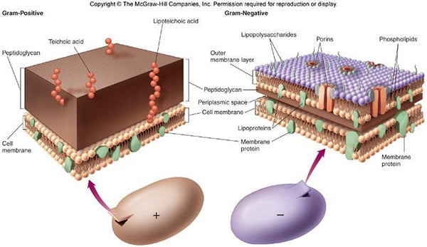
The cell wall is the outermost and non-living part of the cell. Peptidogylcan also called murein. The structural differences between Gram-positive and Gram-negative bacteria cell walls.
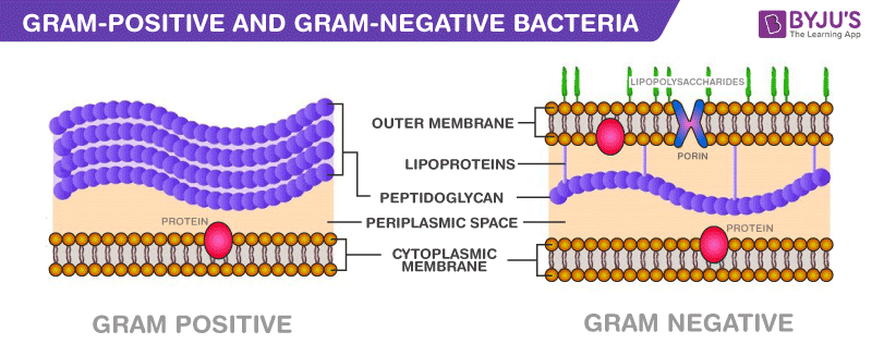
Explain the structural difference between gram-positive and gram-negative bacterial cells that result in the different colors when stained. Located between the plasma membrane and the thin peptidoglycan layer is a gel-like matrix called periplasmic space. Gram positive bacteria have lots of peptidoglycan in their cell wall which allows them to retain crystal violet dye so they stain purple-blue.
There are structural differences between Gram positive and Gram negative bacteria that makes them appear different after Gram staining experiment. Difference Between Gram-Positive and Negative Cell Wall. Include the fluid mosaic model in.
Cell walls are made up of peptidoglycan also known as murein. Thus the two types of bacteria are distinguished by gram staining. Structure of bacterial cytoplasmic membranes.
It is present only in plant cells and also seen in some fungi bacteria and algae. The cell wall of the former kind is thicker peptidoglycan wall whereas that of latter is thinner. Gram-negative bacteria are more resistant against antibodies because their cell wall is impenetrable.
Write an essay about the structural differences between Gram-positive and Gram-negative bacteria cell walls. We are not referring to the gram stain procedure itself Q2. In gram-negative cells there are two structures that need to be mentioned.
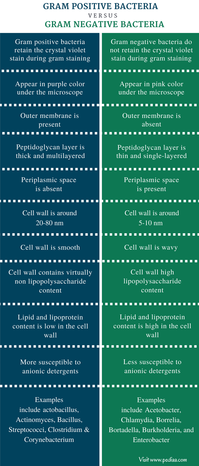
During the gram staining procedure a gram-positive cell retains the purple-colored stain. Cell Wall is single layered straight even less elastic and more rigid and the rigidity of the cell wall is due to the high amount of. Gram negative bacteria have less peptidoglycan in their cell wall so cannot retain crystal violet dye so they stain red-pink.
Cell wall of Gram negative bacteria. The gram-positive bacteria retain the crystal violet colour and stains purple whereas the gram-negative bacteria lose crystal violet and stain red. Whereas the cell wall of gram-negative bacteria is consisting of thin layers of peptidoglycan.
Cell wall is single layered and primarily made up of peptidoglycan. Explain the differences between gram-positive and gram-negative cell walls of bacteria and compare them with the cell walls of archaeobacteria and mycoplasmas. Its 20 to 80 nanometers thick.
The Gram staining technique is dependent on the characteristics of the cell wall. Thickness of peptidoglycan layer teichoic acids outer membrane LPS STUDY. Gram positive bacteria possess the cell wall that is continuous called the sacculus.
Gram positive cell wall retain the primary stain of Gram staining crystal violet and appear purple after alcohol treatment. The cell wall is a thick rigid semi-permeable membrane and has elastic properties that help them to grow in thickness over the period of time. Gram-positive has a large quantity of peptidoglycan and gram-negative have a much smaller amount.
Gram-staining is a differential staining technique that uses a primary stain and a secondary counterstain to distinguish between gram-positive and gram-negative bacteria. Explain how the initiative diddid not address issues of cultural congruency economics and social justice. Describe the structure and functions of the prokaryotic plasma membrane.
Cell wall is made of. Peptidoglycan which is around 80 in gram-positive bacteria whereas in gram-negative bacteria the cell wall is bi-layered wavy uneven. But do not retain the purple colored stain.
Unlike in Gram positive bacteria Gram negative bacteria have an outer membrane layer that is external to the peptidoglycan cell wall. Another difference is gram. The cell wall of gram-positive bacteria is consisting of thick layers of peptidoglycan.
Cell wall is double layered and with an outer membrane outside to Peptidoglycan layer. Cell Wall Structure in Gram Positive Bacteria.
Differences Between Gram Positive And Gram Negative Bacteria
Major Difference Between Gram Positive And Gram Negative Bacteria
Difference Between Gram Positive And Gram Negative Bacteria Definition Cell Wall Structure Characteristics


Comments
Post a Comment